Large-scale, spatially resolved panoramic CRISPR screening in native tissue environments using Perturb-DBiT
What if CRISPR screens could reveal not just which genes matter, but exactly where in a tumor they matter — and how they rewire the entire RNA landscape in situ? That is precisely what Perturb-DBiT now makes possible.
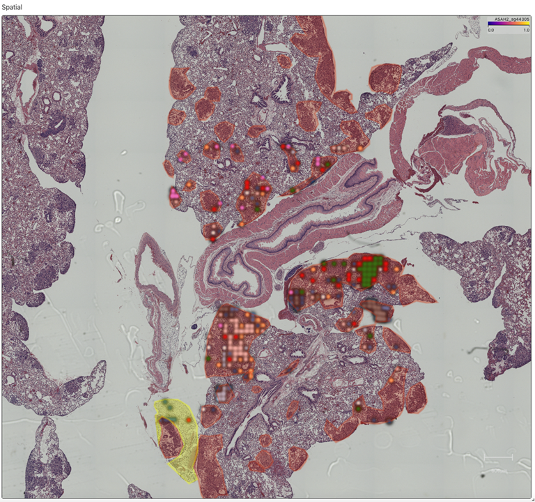
Baysoy, Tian, et al present Perturb-DBiT (perturbation-compatible deterministic barcoding in tissue), a sequencing-based platform that simultaneously co-profiles single guide RNAs (sgRNAs) and spatial total RNA whole transcriptomes on the same intact tissue section. The method was validated across multiple in vivo models and tissue types, including:
- Autochthonous liver cancer genetically engineered mouse models (GEMMs) using a 288-sgRNA tumor suppressor gene library
- A human colorectal cancer (HT29) lung metastatic colonization model using a genome-scale Brunello library of 77,441 sgRNAs
- An immune-competent syngeneic E0771 mouse model using the Brie genome-scale library of 78,637 sgRNAs
- Fresh-frozen and FFPE tissue compatibility, with cross-platform validation against 10x Xenium
Crucially, Perturb-DBiT captures the full RNA repertoire — including lncRNAs, miRNAs, tRNAs, and other noncoding species — alongside protein-coding mRNAs, enabling unprecedented insight into post-transcriptional regulatory responses to genetic perturbation.
Aspect Analytics' Contribution
Aspect Analytics contributed directly to this work by developing a strategy for integrating and visualizing the spatial sgRNA readouts with tissue histology and pathologist annotations using Weave®. The pathologist annotations of the H&E-stained microscopy image were conducted in Weave’s pathology annotation user interface. The location of sgRNA measurements was precisely detected by co-registering its Barcode A and B images to the corresponding brightfield image, using an automatic intensity matching co-registration workflow in Weave. The Perturb-DBiT brightfield image was then co-registered to its serial H&E-stained microscopy image via a manual control point based non-rigid co-registration workflow in Weave. Following this, the sgRNA data was then precisely and directly linked to their histological location. The sgRNA data, histology image and annotations were overlaid and interactively visualized in Weave’s web-based reports.
This collaboration reflects the broader role Aspect Analytics and the Weave platform play in advancing spatial biology: integrating heterogeneous spatial datasets, enabling non-rigid co-registration of consecutive tissue sections, and providing the analytical infrastructure needed to move from raw spatial readouts to interpretable biological insight. The work showcases how Aspect Analytics’ services and Weave's capabilities in multi-modal data integration and high-resolution spatial visualization are essential enablers for next-generation spatial functional genomics.
Please contact us if you are interested in learning more about Weave and our spatial biology services.
Publication Details: Alev Baysoy1,†, Xiaolong Tian1,2,†, Paul Renauer2,†, Feifei Zhang2,†, Zhiliang Bai1, Hao Shi3, Mingyu Yang1, Dingyao Zhang2, Miao Liu1, Haikuo Li1, Bo Tao1, Archibald Enninful1, Yao Lu1, Fu Gao1, Guangchuan Wang2, Wanqiu Zhang4, Thao Tran4, Nathan Heath Patterson4, Jie Sheng5, Shuozhen Bao1, Chuanpeng Dong2, Shan Xin2, Binfan Chen2, Mei Zhong1,6, Sherri Rankin3, Cliff Guy3, Yan Wang3, Jon P. Connelly7, Shondra M. Pruett-Miller7, Daifeng Wang5, Mina Xu8, Mark B. Gerstein2,9, Hongbo Chi3, Sidi Chen2,10,11 & Rong Fan1,8,10,11,12
Large-scale, spatially resolved panoramic CRISPR screening in native tissue environments using Perturb-DBiT
Nature Biotechnology, 2026 · https://doi.org/10.1038/s41587-026-03127-y
AFFILIATIONS
1 Human and Translational Immunology, Yale University School of Medicine, New Haven, CT, USA
2 Department of Genetics, Yale University School of Medicine, New Haven, CT, USA
3 Department of Immunology, St. Jude Children's Research Hospital, Memphis, TN, USA
4 Aspect Analytics NV, Genk, Belgium
5 Department of Biostatistics and Medical Informatics, University of Wisconsin–Madison, Madison, WI, USA
6 Department of Dermatology, Yale University School of Medicine, New Haven, CT, USA
7 Center for Advanced Genome Engineering, St. Jude Children's Research Hospital, Memphis, TN, USA
8 Department of Pathology, Yale University School of Medicine, New Haven, CT, USA
9 Department of Molecular Biophysics and Biochemistry, Yale University, New Haven, CT, USA
10 Yale Stem Cell Center, Yale University School of Medicine, New Haven, CT, USA
11 Yale Cancer Center, Yale University School of Medicine, New Haven, CT, USA
12 Department of Biomedical Engineering, Yale University, New Haven, CT, USA
† These authors contributed equally to this work.