

Integration of Multiple Spatial Omics Modalities Reveals Unique Insights into Molecular Heterogeneity of Prostate Cancer
In this preprint, we integrated spatial transcriptomics, mass spectrometry-based lipidomics, single nucleus RNA-seq and histomorphological information from human prostate cancer patient samples. This provided novel insights into correlating genes and lipids linked to distinct cell populations and histopathological disease states, as well as comparisons across samples in a tissue cohort.
Access publication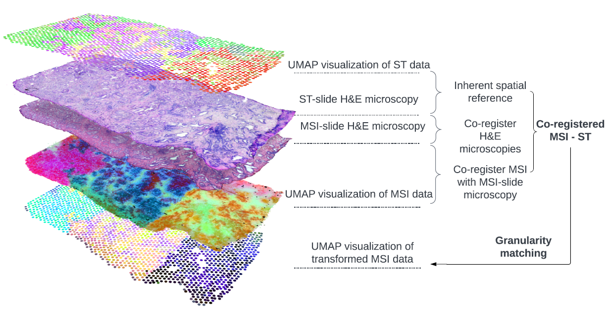
This study introduces our spatial multi-omics integration pipeline (SMOx) designed to improve the alignment and integration of different spatial omics technologies. Unlike previous methods that used simpler strategies such as affine co-registration or weighted nearest neighbor approaches, SMOx incorporates multi-point non-rigid co-registration and Gaussian-weighted granularity matching for more precise data alignment.
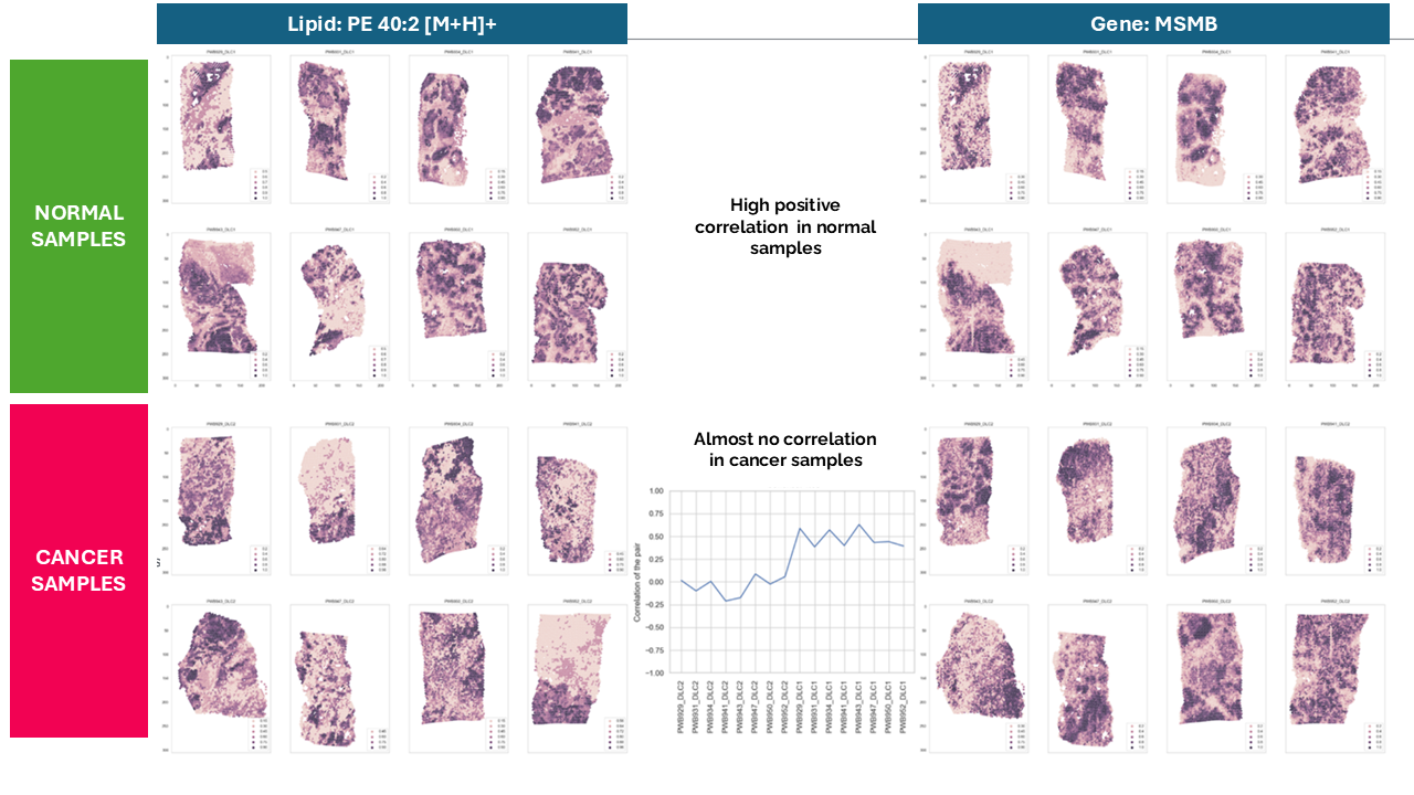
To demonstrate the pipeline’s utility, the team studied human prostate cancer (PCa), a disease known for its morphological and molecular heterogeneity. The researchers applied SMOx to integrate Visium Spatial Transcriptomics (ST), MALDI-2 mass spectrometry imaging for spatial lipidomics (MSI), single-nucleus RNA sequencing (snRNA-seq), and pathology annotations. Integration with the SMox pipeline enabled identification of lipid-gene expression associations tied to specific cell populations and PCa pathological states that are not visible through morphology alone. When applied to a cohort of PCa and matched benign samples, we found that ST and MSI data largely followed histomorphological patterns, and that combining the two analyses allowed us to identify genetic and lipid expression profiles corresponding to different cell populations and tissue states. Our integrated spatial multi-omics approach was able to identify not only new correlating and anti-correlating gene-lipid pairs throughout the cohort, but also the loss of gene-lipid co-expression between benign and PCa samples. These insights have potential applications in molecular pathology, particularly in analyzing ambiguous tissue regions and generating new research hypotheses.
Do not hesitate to contact us if you are interested in learning more about this or have similar workflow needs.
Publication Details: Wanqiu Zhang1,2, Xander Spotbeen3,4, Sebastiaan Vanuytven5,6, Sam Kint4,5, Tassiani Sarretto7, Fabio Socciarelli8, Katy Vandereyken4,5, Jonas Dehairs3, Jakub Idkowiak3,9, David Wouters4,10, Jose Ignacio Alvira Larizgoitia4,10, Gabriele Partel4,10, Alice Ly2, Vincent de Laat3, Maria José Q Mantas2, Thomas Gevaert11, Wout Devlies11,12, Chui Yan Mah13,14,15, Lisa M Butler13,14,15, Massimo Loda8, Steven Joniau11,12, Bart De Moor1, Alejandro Sifrim4,10, Shane R. Ellis7, Thierry Voet4,5, Marc Claesen2, Nico Verbeeck2, Johannes V. Swinnen3,4
Integration of Multiple Spatial Omics Modalities Reveals Unique Insights into Molecular Heterogeneity of Prostate Cancer. doi: https://doi.org/10.1101/2023.08.28.555056.
AFFILIATIONS
1 STADIUS Center for Dynamical Systems, Signal Processing and Data Analytics, Department of Electrical Engineering (ESAT), KU Leuven, Kasteelpark Arenberg 10, 3001
Leuven, Belgium
2 Aspect Analytics NV, C-mine 12, 3600 Genk, Belgium
3 Laboratory of Lipid Metabolism and Cancer, KU Leuven and Leuven Cancer Institute (LKI), Leuven, Belgium
4 KU Leuven Institute for Single Cell Omics (LISCO), KU Leuven, Leuven, Belgium
5 Laboratory of Reproductive Genomics, Department of Human Genetics, KU Leuven, Leuven, Belgium
6 Laboratory of Stem Cells and Cancer, Université Libre de Bruxelles (ULB), Brussels, Belgium
7 Molecular Horizons and School of Chemistry and Molecular Bioscience, University of Wollongong, Northfields Ave, Wollongong, NSW 2522 Australia
8 Department of Pathology and Laboratory Medicine, Weill Cornell Medical College, New York, NY, USA
9 Department of Analytical Chemistry, Faculty of Chemical Technology, University of Pardubice, Pardubice, Czech Republic
10 Laboratory of Multi-omic Integrative Bioinformatics, Department of Human Genetics, KU Leuven, Leuven, Belgium
11 Department of Urology, University Hospitals Leuven, Leuven, Belgium
12 Department of Development and Regeneration, KU Leuven, Leuven, Belgium
13 South Australian Health and Medical Research Institute (SAHMRI), Adelaide, SA 5000, Australia.
14 South Australian Immunogenomics Cancer Institute, University of Adelaide, Adelaide, SA 5005, Australia.
15 Freemasons Centre for Male Health and Wellbeing, University of Adelaide, Adelaide, SA 5005, Australia.

